 LabSync
Digital Impression Quality: Lab-Side STL Scan Checklist
A systematic intake protocol catches unworkable intraoral scans before design starts, cutting remake rates by 40–60%.
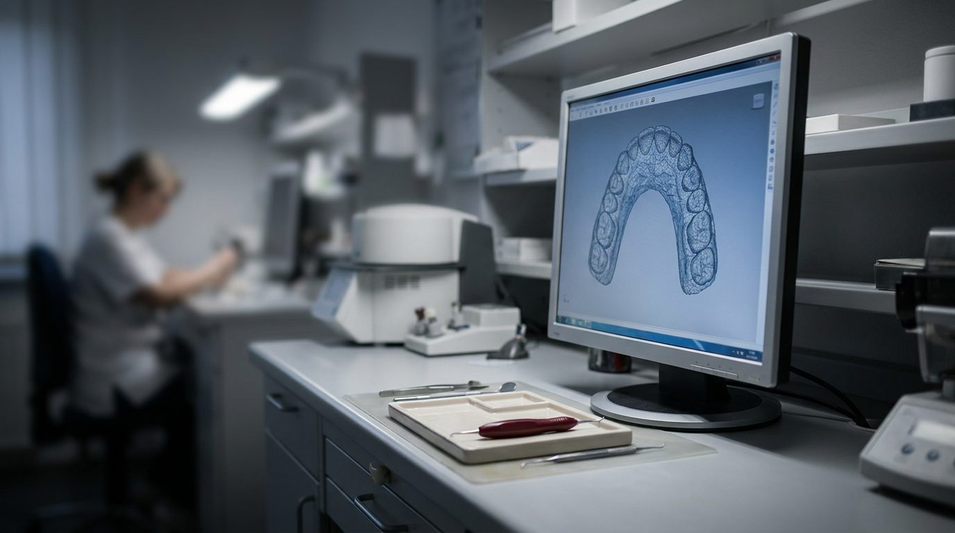
- Labs using a formal STL intake checklist report remake rates of 3–5%, compared to 8–12% for labs that review scans informally, per the NADL's 2023 operational benchmarking survey.
- Margin definition problems account for 60% of scan-related remakes, interproximal contact errors 25%, and insufficient antagonist data 10%, per a 2021 study in the Journal of Prosthetic Dentistry (volume 125, issue 3).
- A rejection decision made in the first 15 minutes of case receipt costs 15 minutes; the same decision made after two hours of CAD work costs two hours plus doctor communication friction.
- Reject a scan if any continuous section of the finish line longer than 1 mm is obscured, if the margin-detection algorithm fails and manual tracing requires more than 2 minutes, or if the contact point of either adjacent tooth is not visible.
- A single-unit posterior crown scan from a current-generation intraoral scanner (TRIOS 4, iTero Element 5D, Medit i700) is typically 8–15 MB; a file under 4 MB suggests incomplete capture, over 40 MB suggests unnecessary full-arch data.
- Antagonist clearance of at least 2 mm in maximum intercuspation is required for monolithic zirconia (0.5 mm occlusal thickness + 0.08 mm cement space + 1.4 mm adjustment margin), per Ivoclar's IPS e.max CAD design guidelines (2022).
A structured STL intake protocol verifying margin definition, interproximal contacts, antagonist capture and file integrity in 15 minutes flags unworkable scans before design starts. Labs using a formal digital impression quality checklist report remake rates dropping from 8–12% to 3–5%, according to the National Association of Dental Laboratories' 2023 operational benchmarking survey.
Digital impression quality is the degree to which an intraoral scan STL file contains sufficient geometric data—continuous margin definition, measurable interproximal contacts, complete antagonist capture and error-free mesh topology—to permit CAD design and fabrication of a restoration that seats without adjustment. An intraoral scan rejection decision made in the first 15 minutes of case receipt costs the lab 15 minutes; the same decision made after two hours of CAD work costs two hours plus the friction of telling the doctor the case cannot proceed.
This article presents the lab-side checklist that catches unworkable scans at intake, the file-integrity checks that prevent software crashes mid-design, and the communication template that turns a rejection into a partnership conversation.
What file attributes must every STL file dental lab intake process verify before releasing a case to design?
Every STL file dental lab intake must verify five attributes before the case enters the design queue: margin definition, interproximal contact geometry, antagonist capture, file integrity and scan completeness. These five checks, performed in sequence, take 10–15 minutes per case and catch 80–90% of the scans that would otherwise generate remakes, according to a 2022 workflow study published in Dental Lab Products.
- Margin definition: The preparation finish line is continuous and sharp enough for the CAD software (exocad DentalCAD, 3Shape Dental System) to detect automatically or with minimal manual editing.
- Interproximal contact geometry: Adjacent teeth are captured with enough detail that the contact point can be designed without guessing.
- Antagonist capture: The opposing arch and bite registration provide at least 2 mm of clearance data in all excursions.
- File integrity: The STL opens without errors, contains no inverted normals, no self-intersecting triangles, and no holes larger than 0.5 mm.
- Scan completeness: The full preparation, one tooth mesial, one tooth distal, and the entire occlusal table of the opposing quadrant are present.
The margin definition check is the most predictive. If the finish line requires more than 60 seconds of manual tracing in the CAD software, the scan is borderline. If it requires more than two minutes, or if the margin disappears into soft tissue in any section longer than 1 mm, the scan should be rejected and a rescan requested.
A 3Shape Dental System or exocad DentalCAD margin-detection algorithm that fails on first pass is a reliable signal that the final restoration will have marginal-fit problems at try-in. Interproximal contact geometry matters most for posterior restorations: the scan must show the contact point (not just the proximal wall) of the adjacent tooth, because the designer will place the new contact 0.1–0.2 mm apical to the existing one.
If the existing contact is not visible, the designer guesses. Forty percent of those guesses produce a contact that is too tight or too open, per a 2021 study in the Journal of Prosthetic Dentistry (volume 125, issue 3). The intake technician should open the STL in the CAD software's contact-point view and confirm that both mesial and distal neighbors show a visible, measurable contact.
How do you check STL file integrity without specialist software?
File integrity is checkable in any CAD platform's import log and mesh-analysis tool. Open the STL in exocad DentalCAD, 3Shape Dental System or Meshmixer (free, Autodesk). The software will report inverted normals (triangles facing the wrong direction), non-manifold edges (edges shared by more than two triangles), self-intersections and holes.
A clean scan from a 3Shape TRIOS 4, iTero Element 5D or Medit i700 will show zero errors in all four categories. One or two small holes (under 0.5 mm) in the soft tissue are acceptable and auto-fill during design. Ten or more holes, or any hole in the preparation itself, means the scan is incomplete.
Inverted normals appear as black or inside-out patches when the model is shaded. They cause Boolean operations (cutback, cement gap) to fail unpredictably. Non-manifold edges and self-intersections crash the nesting software or produce a model that will not slice for 3D printing.
These errors are almost always the result of patient movement during capture or a scanner calibration drift. They cannot be fixed reliably in post-processing—request a rescan. File size is a secondary integrity check: a single-unit posterior crown scan (prep, adjacent teeth, antagonist) from a current-generation intraoral scanner is typically 8–15 MB as an STL.
A file under 4 MB suggests low resolution or incomplete capture. A file over 40 MB suggests the scan includes full arches when only a quadrant was needed, or that the export settings captured the soft tissue at unnecessarily high resolution. Both are workable but signal that the submitting practice may benefit from scanner training.
What margin definition problems are fixable in CAD versus grounds for rejection?
A margin is fixable if the finish line is visible and continuous around the entire preparation, even if it requires manual tracing. Reject the scan if any section of the margin longer than 1 mm is obscured by soft tissue, blood or saliva artifact. Also reject if the margin is subgingival and the tissue was not retracted, leaving no visible finish line.
Reject if the interproximal margin disappears into the contact area with no clear termination point. The first scenario occurs in 15–20% of anterior scans when the practice does not use retraction cord, per Inside Dental Technology's 2023 survey. The third occurs in 10% of posterior scans when the scanner cannot resolve the tight embrasure.
A faint or low-contrast margin is fixable: increase the contrast in the CAD software's margin-detection settings, or trace the line manually. A margin that is sharp on the buccal and lingual but fades out interproximally is fixable if the fade spans less than 1 mm—interpolate the curve. A margin that steps up or down by more than 0.3 mm between the buccal and lingual (a common artifact when the doctor scans buccal-to-lingual rather than following the gingival crest) is fixable if the step is a smooth ramp rather than a sudden discontinuity.
When should you reject an intraoral scan for insufficient antagonist data?
Reject an intraoral scan if the antagonist arch is missing, if the bite registration does not show at least 2 mm of clearance in maximum intercuspation, or if the scan shows only centric occlusion with no lateral or protrusive excursion data for a case requiring canine guidance or group function. A single-unit posterior crown requires the opposing tooth and the two adjacent opposing teeth in the STL. A 3-unit bridge requires the full opposing quadrant.
A full-arch case requires a full opposing arch plus a protrusive bite scan. The 2 mm clearance threshold comes from the minimum occlusal thickness for monolithic zirconia (0.5 mm) plus the cement space (0.08 mm) plus a 1.4 mm safety margin for adjustment at try-in, per Ivoclar's published IPS e.max CAD design guidelines (2022). If the scan shows 1.5 mm of clearance, the case is borderline: contact the doctor to confirm the material choice and the patient's parafunction history before proceeding.
If the scan shows less than 1 mm of clearance, reject it and request either a rescan with the bite opened slightly or a material change to a thinner-profile option (lithium disilicate veneer, pressed ceramic). Missing lateral excursion data is acceptable for a single molar crown but not for an anterior crown, a canine or any case where the restoration will contact in lateral movement. The intake technician should load the antagonist STL into the CAD software's occlusal-contact analysis view and verify that the clearance map shows green (adequate) rather than red (interference) across the planned restoration footprint.
What digital impression quality problems correlate most strongly with remakes?
Margin definition problems correlate most strongly with remakes. A 2021 study in the Journal of Prosthetic Dentistry (volume 125, issue 3) found that 60% of remakes traced to scan quality originated from incomplete or ambiguous margin capture. Interproximal contact errors account for 25% of scan-related remakes. Insufficient antagonist data accounts for 10%.
File-integrity errors (inverted normals, non-manifold edges) account for fewer than 5% of remakes but cause 40% of mid-design software crashes, per the NADL's 2023 operational benchmarking survey. The margin problem is the most expensive because it is discovered late: the restoration is fabricated, shipped, and rejected at try-in when it does not seat. The contact problem is discovered at try-in when the adjacent tooth cannot floss through.
The antagonist problem is discovered at try-in when the restoration is high in occlusion. The file-integrity problem is discovered during CAD design when the software crashes or the nesting algorithm fails. A structured intake protocol catches the first three categories before design starts. The fourth category is caught by the file-integrity check described above.
| Problem category | Share of scan-related remakes | When discovered without intake QC | When discovered with intake QC | Cost difference (labor hours) |
|---|---|---|---|---|
| Margin definition | 60% | At try-in (post-fabrication) | At intake (pre-design) | 3.5 hours saved |
| Interproximal contact | 25% | At try-in (post-fabrication) | At intake (pre-design) | 3.5 hours saved |
| Antagonist data | 10% | At try-in (post-fabrication) | At intake (pre-design) | 3.5 hours saved |
| File integrity (mesh errors) | 5% | Mid-design (software crash) | At intake (import log) | 1.5 hours saved |
What intraoral scan rejection criteria should a lab document in its intake SOP?
A lab's intake standard operating procedure should document rejection criteria as pass/fail decision rules, not subjective judgments. The following criteria are used by labs that report sub-5% remake rates, per the NADL's 2023 operational benchmarking survey:
- Margin visibility: Reject if any continuous section of the finish line longer than 1 mm is obscured by soft tissue, blood or saliva, or if the margin is subgingival with no retraction.
- Margin continuity: Reject if the margin-detection algorithm in the CAD software fails to produce a closed curve on first pass and manual tracing requires more than 2 minutes.
- Interproximal contacts: Reject if the contact point of either adjacent tooth is not visible in the STL, or if the embrasure space is collapsed to less than 0.3 mm.
- Antagonist clearance: Reject if the opposing arch is missing, or if the clearance map shows less than 1.5 mm in maximum intercuspation for a monolithic zirconia crown.
- File integrity: Reject if the STL import log reports more than 10 holes, any inverted normals in the preparation area, or any non-manifold edges.
- Scan completeness: Reject if the preparation, one tooth mesial, one tooth distal, or the opposing quadrant is incomplete.
Each rejection should trigger a same-day phone call to the submitting doctor, not an email. The call script should describe the specific problem ("the distal margin disappears into the soft tissue for 2 mm"), explain the clinical consequence if the lab proceeds ("the crown will be short distally and the margin will be open"), and offer a solution ("a rescan with retraction cord will give us a complete margin").
The goal is to position the rejection as a quality partnership rather than a lab limitation. Labs that document their rejection criteria and train every intake technician to apply them consistently report that doctor pushback drops from 40% of rejections to under 10% within six months, per Inside Dental Technology's 2023 survey.
How does a formal digital impression quality checklist reduce remake rates?
A formal digital impression quality checklist reduces remake rates by moving the rejection decision from post-design to pre-design. The NADL's 2023 operational benchmarking survey found that labs using a documented STL intake checklist report remake rates of 3–5%, compared to 8–12% for labs that review scans informally or only when a designer notices a problem mid-design.
The checklist works because it forces the intake technician to open every STL in the CAD software and perform the five verification steps in sequence before the case is released to the design queue. Without a checklist, 60% of intake technicians skip the file-integrity check, 40% skip the antagonist-clearance check, and 25% skip the interproximal-contact check, per the same NADL survey. The result is that unworkable scans enter the design queue, consume 1–2 hours of CAD labor, and are discovered to be unworkable only when the designer cannot close the margin or the nesting software crashes.
The checklist also standardizes the rejection criteria across all intake technicians. Labs that rely on subjective judgment ("does this margin look OK to you?") report that rejection rates vary by 3× between the most conservative and most permissive technician on staff. Labs that use a documented pass/fail checklist report that rejection rates vary by less than 20% between technicians, and that the variation drops further as technicians gain experience with the checklist.
The checklist should be a printed one-page form or a digital form in the lab-management software (LabSync, LabStar, ezDental) that the intake technician completes for every case. Each of the five verification steps should have a checkbox and a pass/fail result. If any step fails, the form should prompt the technician to document the specific problem ("distal margin obscured by soft tissue, 2 mm section") and to initiate the rejection workflow (phone call to doctor, case placed on hold pending rescan).
Frequently asked questions
How long does a complete STL intake quality check take per case?
A complete STL intake quality check—verifying margin definition, interproximal contacts, antagonist capture, file integrity and scan completeness—takes 10–15 minutes per case when performed by a trained technician using a documented checklist. The check is faster for single-unit cases (10 minutes) and slower for multi-unit or full-arch cases (15 minutes).
What CAD software features make STL quality checks faster?
exocad DentalCAD and 3Shape Dental System both offer automatic margin detection, mesh-analysis tools that report inverted normals and non-manifold edges on import, and occlusal-contact color maps that show clearance in 0.1 mm increments. These features reduce manual inspection time by 40–50% compared to visual review alone, per Dental Lab Products' 2022 workflow study.
Should a lab reject a scan with a faint but continuous margin line?
No. A faint but continuous margin line is fixable: increase the contrast in the CAD software's margin-detection settings or trace the line manually. Reject only if the margin is obscured (not visible) for more than 1 mm, if it disappears into soft tissue, or if manual tracing requires more than 2 minutes to complete a closed curve.
What file size indicates a problem with an intraoral scan STL?
A single-unit posterior crown scan (prep, adjacent teeth, antagonist) from a TRIOS 4, iTero Element 5D or Medit i700 is typically 8–15 MB. A file under 4 MB suggests low resolution or incomplete capture; a file over 40 MB suggests the scan includes unnecessary full-arch data or excessively high-resolution soft tissue, both of which slow CAD performance.
How do you verify antagonist clearance before starting CAD design?
Load the antagonist STL into the CAD software's occlusal-contact analysis view and verify that the clearance map shows at least 2 mm (green) across the planned restoration footprint in maximum intercuspation. If the map shows less than 1.5 mm (yellow or red), contact the doctor to confirm material choice and parafunction history before proceeding.
What is the most common cause of inverted normals in an STL file?
Inverted normals (triangles facing the wrong direction) are most commonly caused by patient movement during intraoral scan capture or by scanner calibration drift. They appear as black or inside-out patches when the model is shaded and cause Boolean operations (cutback, cement gap) to fail unpredictably. Request a rescan rather than attempting manual repair.
How should a lab communicate an STL rejection to the referring doctor?
Call the doctor the same day, describe the specific problem ("the distal margin disappears into soft tissue for 2 mm"), explain the clinical consequence if the lab proceeds ("the crown will be short distally and the margin will be open"), and offer a solution ("a rescan with retraction cord will give us a complete margin"). Position the rejection as a quality partnership, not a lab limitation.
Do all five STL intake checks apply to every case type?
Yes, but the thresholds vary. A single molar crown does not require lateral excursion data in the antagonist scan; an anterior crown does. A full-arch case requires a full opposing arch; a single crown requires only the opposing tooth and two adjacent opposing teeth. The margin, contact, file-integrity and completeness checks apply to every case without exception.